:: a brief history of CT
:: contents
introductionscientific background and early technical development
clinical acceptance and early commercial development
further development
current use of CT
CT and other imaging modalities
further reading (and viewing)
timeline 1895-2011
introduction
The first clinical CT scan on a patient took place on 1st October 1971 at Atkinson Morley's Hospital, in London, England. The patient, a lady with a suspected frontal lobe tumour, was scanned with a prototype scanner, developed by Godfrey Hounsfield and his team at EMI Central Research Laboratories in Hayes, west London. The scanner produced an image with an 80 x 80 matrix, taking about 5 minutes for each scan, with a similar time required to process the image data. Current CT scanners can produce images with an 1024 x 1024 matrix, acquiring data for a slice in less than 0.3 seconds, and are an integral part of a modern hospital's imaging resources.
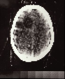
The first clinical scan: Atkinson Morley's Hospital, October 1971
scientific background and early technical development
Computed Tomography (CT) builds on developments in two fields - X-ray imaging and computing. X-rays were discovered in 1895 and within a few years were an established medical tool. By the 1930s, tomography was being developed, enabling the visualisation of sections through a body. By the 1960s, several researchers had worked independently on cross-sectional imaging, culminating in Hounsfield's work at EMI developing computed tomography (CT) for the EMI Scanner. This device relied on the reconstruction of image data by computer, the data being acquired from multiple X-ray transmissions through the object under investigation.
clinical acceptance and early commercial development
Following the first clinical scan in 1971, the patient with the suspected frontal lobe tumour was operated on. The surgeon performing the operation is reported to have remarked that "it looks exactly like the picture" shown above. The successful demonstration of the worth of CT lead to further demonstration systems being installed in London, Manchester and Glasgow in the UK and at the Mayo Clinic and Massachusetts General Hospital in the USA. The first clinical scan in the USA was performed at the Mayo Clinic in 1973.
Papers relating to the first commercial scanner to be installed, the EMI Head CT Scanner at the Manchester Royal Infirmary, and of later EMI scanners installed in Manchester, are held in the Papers of Ian Isherwood collection at the John Rylands University Library, Manchester.
By 1975, EMI were marketing a body scanner, the CT5000, the first of which was installed at Northwick Park Hospital in London. The first body scanner in the USA was installed at the Mallinkrodt Institute and had its first clinical use in October 1975. By this time scan time had been reduced to 20 seconds, for a 320 x 320 image matrix.
The mid-1970s were a time of rapid development in CT: 1976 saw 17 companies offering scanners, with scan times down to 5 seconds in some cases. By 1978, there was an installed base of around 200 scanners in the USA, image matrix sizes were up to 512 x 512 and some models of scanner had the capability of ECG-triggered scans. It was also around this time that the ImPACT group was established at St George's Hospital in London, with funding from the UK Department of Health and tasked with evaluating CT scanners for the UK NHS.
By the end of the 1970s the importance of CT scanning to medicine was clear: Hounsfield and McCormack received the Nobel Prize for Medicine in 1979, for the independent work on developing the theory and technology of CT scanning, and in 1981 Hounsfield received a knighthood for his work.
further development
The 1980s saw incremental development of CT scanner technology: shorter scan times and increased matrix sizes, until by the late 1980s scan times were down to only 3 seconds and matrix sizes were up to 1024 x 1024. Development continued through the 1990s, with the introduction of spiral (continuous) scanning in the early 1990s and the development of multi-slice scanners, with 4-slice scanners and 0.5 second scan times being 'state-of-the-art' by the end of the century.
current use of CT
Development of CT scanner technology continued through the early years of the 21st century, particularly with multi-slice scanners. At the time of writing, high-end scanners were offering up to 320 slices, dual-source and dual-energy x-ray sources and iterative reconstruction techniques.
CT and other imaging modalities
CT is not the only cross-sectional modality in use in medical imaging today. Magnetic Resonance Imaging (MRI) was introduced commercially in 1980, a few years after the introduction of the PET scanner. Each device has its advantages: PET gives functional information whereas MRI and CT give structural information. The PET/CT scanner, which combines information from a PET scan and a CT scan in a single device, was introduced in 2000.
further reading (and viewing)
- Beckmann, EC. CT scanning the early days. BJR 2006; 79:5-8 link
- Hounsfield, GN. Computerised transverse axial scanning (tomography): Part 1. Description of system. BJR 1973;46:1016-22
- Ambrose, J. Computerised transverse axial scanning (tomography): Part 2. Clinical application. BJR 1973;46:1023-47
- Perry, BJ and Bridges, C. Computerised transverse axial scanning (tomography): Part 3. Radiation dose considerations. BJR 1973;46:1048-1051
- "Godfrey N. Hounsfield - Autobiography". Nobelprize.org. 16 Mar 2011 http://nobelprize.org/nobel_prizes/medicine/laureates/1979/hounsfield-autobio.html link
- Godfrey N Hounsfield. Computed Medical Imaging. Nobel Prize lecture, 8th December 1979 link
- Allan MacLeod Cormack. Early Two-Dimensional Reconstruction and Recent Topics Stemming From It. Novel Prize Lecture, 8th December 1979 link
- Webb, S. From The Watching Of Shadows. Adam Hilger, Bristol, 1990
- Hendee, WR. History of Cross Sectional Imaging. Radiographics 1989; (6):1155-1180
- How a CT Scanner works, by Vanderbilt Biomedical Engineers: a musical(?) explanation on YouTube
ImPACT presentations:
- CT Scanner Development
- Early CT scanner dose notebooks
- Basic Principles of CT
- Other ImPACT presentations
Timeline
The timeline below gives key events in the history of CT and related medical imaging technology.
1895
- Wilhelm Conrad Röntgen discovers x-rays in Würzburg, Bavaria, Germany

Herr Professor Dr & Mrs Röntgen, December 1895
1896
- F.H. Williams takes the first successful chest x-ray, in Boston, Massachusetts
- Carl Schleussner, working with Wilhelm Röntgen, develops the photographic X-ray plate, in Frankfurt am Main, Germany
- First X-ray department established, at Glasgow Royal Infirmary, Scotland, UK
1902
- first radiation-induced cancer reported, of the skin
1913
- William David Coolidge invents the hot cathode x-ray tube

from the Cathode Ray Tube Site
1917
- Johann Radon demonstrates that the image of a 3-dimensional object can be reconstructed from an infinite number of 2-dimensional projections of the object, providing the mathematical basis for CT image construction (Vienna, Austria)
from Slice Reconstruction page at University of Edinburgh School of Informatics
1921
- André Bocage develops focal-plane tomography (Paris, France)
1930 - 1931
- Alessandro Vallebona develops 'stratigraphy' (Genova, Italy); Bernard Ziedes des Plantes develops 'planigraphy' (Utrecht, Netherlands): both forms of tomography
1937
- William Watson patents axial transverse tomography and obtains the first radiographic images using this technique (London, UK)
1940
- Gabriel Frank patents back-projection (Budapest, Hungary)
1961
- William Oldendorf builds a model tomographic scanner that utilises the techniques later developed independently by Hounsfield and Cormack (Los Angeles, USA)
- first PET scanner demonstrated by James Robertson and associates (New York, USA)
1963 & 1964
- Allan Cormack publishes in the Journal of Applied Physics a theoretical analysis and the results from experimental scanner using a computer to reconstruct cross-sectional images from data (Medford, Massachusetts, USA)
1966
- David Kuhl, John Hale and Walter Eaton publish a paper with the transmission images of a subject's thorax, using an external radiation source. The first cross-sectional images of a human subject (Philadephia, USA)
1968
- Godfrey Hounsfield's original project proposal at EMI (London, UK)
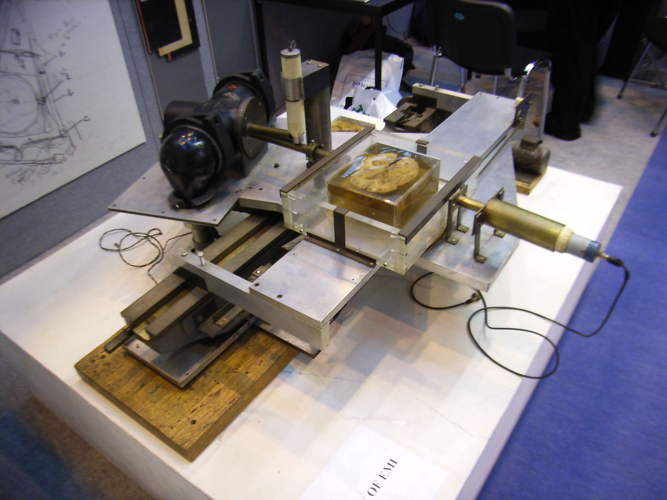
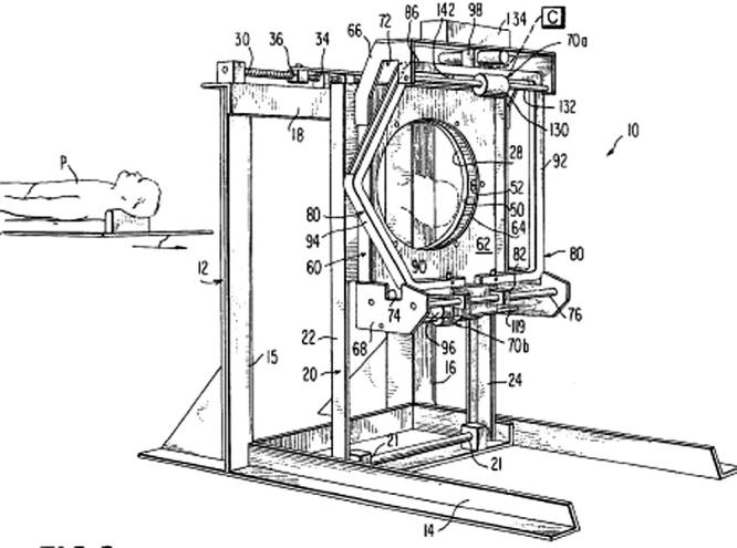
Hounsfield's prototype CT scanner
1969
- Hounsfield meets with Dr James Ambrose and Dr John Perry of St George's Hospital at the suggestion of the UK Department of Health (London, UK)
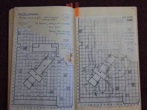
Pages from Dr Perry's notebooks on the first EMI scanner
1971
- Hounsfield lectures on CT of animals (Amsterdam, The Netherlands)
- first patient (head scan), under the supervision of Hounsfield and Ambrose, at Atkinson Morley's Hospital, using prototype EMI head scanner (London, UK)
The first clinical scan, 1st October 1971
1972
- Hounsfield & Ambrose lecture, BIR annual conference (London, UK)
- Godfrey Hounsfield & James Bull lecture, New York, showing first clinical CT images (New York, USA)
- The first CT scanner demonstration in the United States, at the Mayo Clinic (Rochester, MN, USA)
1973
- 320 x 320 image matrix
- EMI awarded the Queen's Award for Technological Innovation for the 'EMI scanner' (London, UK)
- first clinical patient scan in USA, at the Mayo Clinic (Rochester, MN, USA)
- Robert Ledley designs ACTA, a whole body CT scanner, later marketed by Pfizer (Washington, DC, USA)
from Sittig et al, quoting Ledley's patent application
1974
- first body CT scan (of Hounsfield) in a prototype of the EMI body scanner (London, UK)
- 35 EMI head scanners installed worldwide; 60 more on order
1975
- Prototype of the EMI CT1010 body scanner installed at Atkinson Morley's Hospital (London, UK)
- First commercial PET scanner installed (Los Angeles, USA)
- first scans from EMI body scanner shown, at first International Conference on CT (Bermuda)
- research body scanner installed at Northwick Park Hospital (London, UK)
- first body scan in USA, under the direction of Dr Ron Evans at the Mallinkrodt Institute (St Louis, USA)
- 'Second generation' Ohio Nuclear Delta CT scanners are marketed; GE 'third generation' CT scanners are marketed
1976
- 5 second scan time for an image
- Seventeen companies now offering 'third generation' CT scanners
- patent filed for scanning focus system ("flying focal spot")
- 650 scanners now installed worldwide; 450 supplied by EMI
1977
- First MRI body scan on humans, by Raymond Vahan Damadian et al (Nottingham, UK)
- ImPACT established at St George's on short-term funding (London, UK)

1978
- 512 x 512 image matrix
- 200 scanners sold in USA
- ECG-synchronised CT scanning
1979
- Hounsfield and Cormack jointly awarded the Nobel Prize (Stockholm, Sweden)
- Around 1000 scanners in operation worldwide
- EMI sold to Thorn
1980
- Thorn-EMI sells its CT scanner business to GE
- FONAR markets the first commercial MRI scanner (Melville, NY, USA)
1981
- 3 second scan time for an image
- Godfrey Hounsfield knighted (London, UK)
1983
- 800 CT scanners sold in USA
1985
- 1 second scan times
- 'Superfast CT' (Electron Beam Tomography) is developed by Douglas Boyd (San Francisco, USA)
1987
- 1024 x 1024 image matrix
1989
- First spiral (helical) CT, manufactured by Siemens, enters the market (Erlangen, Germany)
1992
- Elscint CT Twin, first modern multi-slice scanner; sub-millimetre slices (Haifa, Israel)

Elscint CT Twin
1994
- 0.75 second scan time
1998
- 4-slice scanners
- 0.5 second scan time
1999
- PET/CT developed, developed by Dr David Townsend and Dr Ron Nutt (Pittsburgh, USA)

Dr Ronald Nutt and Dr David W Townsend, from the IEEE website
2002
- 8- and 16-slice scanners introduced
2007
- 72 million CT scans performed in USA
2011
- Closure of ImPACT, the UK CT scanner evaluation centre, after more than a quarter of a century at St George's Hospital, London (29th September 2011)
- 40th anniversary of the world's first clinical CT scan, undertaken at Atkinson Morley's Hospital, London, UK (1st October 1971)