:: patient's information and guide to ct scanning
This section of the web site has been compiled to answer a number of common questions about CT scanners, and CT examinations.
If you have any other questions, please try some of the excellent information provided by repuatable groups, such as the RCR or HPA and talk to your health care professional
- What is a CT Scanner?
- Why do I need a CT scan rather than an ordinary x-ray?
- What does a CT scanner look like?
- What parts of the body is a CT scanner used for?
- What type of problems can CT scanning diagnose?
- Does a CT scan hurt?
- What is a contrast CT scan?
- How long does a CT scan take?
- Who carries out CT scans?
- How does a CT scanner work?
- What are x-rays?
- What is the radiation dose from a CT scan?
What is a CT Scanner?
A CT or CAT (short for Computed Tomography or Computed Axial Tomography) scanner is a piece of medical imaging equipment that uses x-rays to create images of 'slices' through the body (see figure 1).
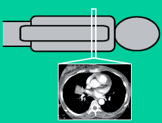
Figure 1: CT image slice
Why do I need a CT scan rather than an ordinary x-ray?
Because of the way CT images are produced, they have advantages over normal x-ray images in distinguishing between different types of soft tissue (e.g. the liver and kidneys, marked in blue and orange in figure 2).
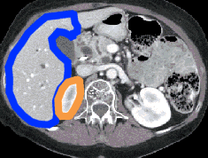
Figure 2: Good contrast between different organs
In addition, multiple CT images can be combined to produce a three-dimensional display, which can be presented in a variety of novel ways (figure 3), and can provide extra information.
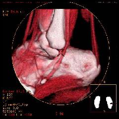
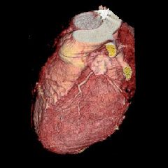
Figure 3a and 3b: Three dimensional reconstructions of CT images of bone and
muscle in the heel and exterior of the heart
What does a CT scanner look like?
A CT scanner looks like a large doughnut shaped gantry, together with a table that moves the patient in and out (figure 4). In addition, there is a computer system that is used to plan and display the images the scanner produces.
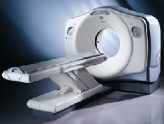
Figure 4: A typical CT scanner
What parts of the body is a CT scanner used for?
CT scanners can be used to image all parts of the body. Some areas which CT scanning is particularly good for include:
- Internal organs within the abdomen and chest, e.g. liver, kidneys, pancreas, intestines, lungs
- Bone imaging, for orthopaedic examinations
- Brain imaging
- Vascular imaging, examining blood flow to different parts of the body
What type of problems can CT scanning diagnose?
Because the CT scanner can be used to image the whole body, there are a huge range of potential uses. Some of the more common ones include:
- Diagnosis and staging of a wide range of cancers
- Assessing organ and bone damage to trauma (accident) victims
- Planning radiotherapy treatment regimes
- Assessment of vasular (blood flow) diseases
- Screening for and assessing cardiac (heart) disease
- Assessment of injury and disease to bones, particularly in the spine
- Bone mineral densitometry for the assessment of osteoporosis
- Guiding biopsy procedures for taking tissue samples
Does a CT scan hurt?
No. Patients are asked to lie still while the scan takes place, and there will be a whirring noise caused by the movement of the parts of the scanner within the gantry. You may need an injection of a special contrast agent during the course of the scan.
What is a contrast CT scan?
For some types of CT scan, a 'contrast agent' is used to make certain parts of the patient more visible to x-rays. The contrast agent is in the form of a liquid that is injected into the patient prior to scanning. This liquid has iodine in it, and shows up well in CT scans. This sort of examination is often performed as a multi-phase study, where two or more sets of images of the organ are taken, e.g. before and after the injection of the contrast agent. The differences between these images can give the radiologist valuable information.
How long does a CT scan take?
The total length of a CT scan procedure can be between approximately 20 minutes to 1 hour. The actual scanning itself takes between a few seconds and a few minutes, depending upon the examination, so most of the procedure time is in preparation and confirmation that the images are of sufficient quality.
Who carries out CT scans?
A CT scan is generally conducted by a radiographer or technologist, under the guidance of a radiologist. The radiologist will also examine and report upon the results of the scan.
How does a CT scanner work?
Inside the doughnut shaped gantry of a CT scanner, there is an x-ray tube. It rotates around the patient, taking hundreds of x-ray pictures of a thin section of the body. This data can be combined mathematically to produce an image through the patient.
What are x-rays?
X-rays are a form of electromagnetic radiation. Other forms of electromagnetic radiation include visible light, radio waves and microwaves. The difference between these different types lies in the energy of the radiation, and their interactions with matter. Visible light has enough energy to penetrate through a thin piece of card, whereas x-rays are more energetic, and can be 'shined' through the body to form an image on a detector on the far side.
What is the radiation dose from a CT scan?
X-rays have the power to do potential damage to the body, as they are a form of ionising radiation. Most scientists think that there is no such thing as a 'safe' dose of radiation, so the exposure of the body to x-rays should be kept to a minimum. Doses from radiation are measured in 'milliSieverts', or mSv. Doses from head, abdomen and chest CT examinations are typically around 1, 5 and 8 mSv respectively. Although this is fairly high in comparison with other doses from x-ray examinations, the images provided by a CT scan can provide more or better information.
In all cases, the clinician who refers you for a CT scan will ensure that the benefits provided by the information in the examination outweigh the risks associated with the radiation dose.