:: rsna 2001: impact on what's new in ct
This article was produced for the quarterly publication 'Diagnostic Imaging Review', printed and published by the Medical Devices Agency (now absorbed into the Medicines and Healthcare Products Regulatory Agency).
Introduction
2001 turned out to be yet another exciting year for CT scanning at RSNA. The prototype eight- and sixteen-slice scanners that appeared at last year's show are now genuine products, although most will not appear in clinical use until the middle of next year. One of the implications of going beyond four slices per rotation is the necessity to design new detector arrays. In addition, the data interpolation and reconstruction algorithms used for four-slice scanners do not extend particularly well to eight slices, so new techniques have had to be introduced. Faster gantry rotation times are also available on some scanners.
Single- and dual-slice scanners have not shown as much progress as their multi-slice counterparts, but some advances have occurred. In particular, a trickle-down of technology and applications means these scanners can perform more advanced examinations than before.
A number of specialised clinical applications featured heavily at RSNA, including colonoscopy, angiography, lung imaging, perfusion imaging and cardiac (particularly functional cardiac imaging). These applications benefit from the capabilities of multi-slice scanning. The trend to more slices should improve them further. All the scanner manufacturers have software packages covering these areas. Some require special scan acquisition modes and others the formidable processing and graphical capabilities of modern workstations to become usable on a routine basis.
New scanners
When four-slice scanners were first introduced to the market at RSNA 1998, the expectation was that they would be purchased mainly by university and teaching hospitals. The market for CT scanners in the UK is different from most others, as purchases tend to be upper range scanners. Nearly all scanners currently being purchased for UK hospitals are four-slice models. This is due to intense competition between the manufacturers driving the prices down, as well as the fast pace of development in CT scanning, which seems to show no signs of slowing.
Last year at RSNA, Marconi and Siemens featured their sixteen-slice scanners of the future. GE showed images from their first clinical eight-slice scanner, and Toshiba indicated that they would soon have a sixteen-slice model available. This year, these plans are more clearly defined. The manufacturers have indicated that, by the second half of 2002, they should all have an eight- or sixteen-slice scanner available for purchase and installation. Some of the relevant details of the scanners are shown in Table 1. The detector layouts for these scanners are illustrated in Figure 1, which shows the arrangement of elements along the patient's long axis (the z-axis). As with four-slice scanners, the output from multiple detectors can be combined in order to vary the slice thickness.
| Manufacturer | Scanner | Fastest rot. time (s) | Slices per rotation | Total detector length (mm) | Collimations (mm) |
|---|---|---|---|---|---|
| GE | LightSpeed Ultra | 0.5 | 8 | 20 | 8 x 1.25, 8 x 2.5 |
| GE | LightSpeed 16 | 0.5 | 16 | 20 | 16 x 0.63, 16 x 1.25 |
| Philips | Mx8000 Infinite | 0.4 | 16 | 24 | 16 x 0.75, 16 x 1.5 |
| Siemens | Sensation 16 | 0.4 | 16 | 24 | 16 x 0.75, 16 x 1.5 |
| Toshiba | Aquilion 16 | 0.5 | 16 | 32 | 16 x 0.5, 16 x 1, 16 x 2 |
Table 1: Eight- and sixteen-slice scanner specification
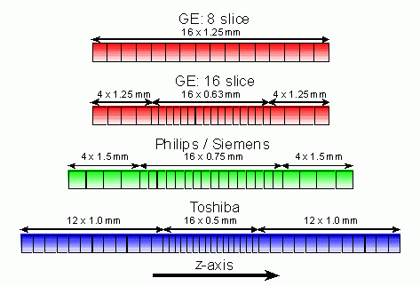
Figure 1: Detector arrangements for eight- and sixteen-slice
GE is the first company to have installations of a scanner with more than four slices, with the LightSpeed Ultra. This is a development of the existing LightSpeed Plus and uses the same detector array, but the number of channels of data has been increased from four to eight. This enables either 8 x 1.25 or 8 x 2.5 mm acquisitions, giving a coverage of up to 20 mm per rotation. GE has more than 150 clinical installations of the LightSpeed Ultra worldwide, and a number of scientific papers in the meeting featured work using this scanner. As with the LightSpeed Plus, the LightSpeed Ultra allows only certain optimised helical pitches in clinical use. For the Ultra, these are 0.625, 0.875, 1.35 and 1.675, which is equivalent to a pitch of 5, 7, 10.8 and 13.4 when the table travel is quoted relative to the detector width. GE is also working on the development of a sixteen-slice scanner (as yet unnamed), and showed some initial images from the first clinical installation. This will also have a 20 mm detector array, but in a configuration that is capable of acquiring 16 x 0.63 or 16 x 1.25 mm slices per rotation.
Philips Medical Systems has recently acquired Marconi Medical Systems, so the Marconi Mx8000 range of multi-slice scanners is now known as the Philips Mx8000. The newest scanner in the Mx8000 range is the sixteen-slice Mx8000 Infinite, which has a 24 mm detector and can acquire 16 x 0.75 or 16 x 1.5 mm slices per rotation. In addition, the fastest gantry rotation speed has been improved from 0.5 to 0.4 seconds, which will allow acquisition at a rate of up to 40 full rotation data sets per second. The Mx8000 Infinite should be available from May 2002. Philips' Secura Trueview four-slice scanner, which was announced at last year's RSNA, will not now be available, although aspects of the technology used in its development will probably appear in the Mx8000 range. For example, the COBRA (Cone Beam Reconstruction Algorithm) used on the Mx8000 Infinite is a development of Philips' n-pi reconstruction methods.
Siemens' newest scanner is the Somatom Sensation 16. It also uses a 24 mm detector, allowing 16 x 0.75 and 16 x 1.5 mm slices per rotation. Itfeatures scan times as fast as 0.4 s per rotation. This is particularly useful for cardiac imaging, where ECG gating techniques can be used to improve the effective scan time. The Sensation 16 has a built-in ECG monitor and the ECG cycle is displayed on the scanner gantry. It should be available in the middle of 2002. Siemens' Somatom Volume Zoom four-slice scanner range has been renamed Somatom Sensation 4.
Toshiba showed its sixteen-slice scanner, the Aquilion 16, which features an updated version of its multi-slice detector. The new detector is also 32 mm long in the z-axis, and can be collimated to provide 16 x 0.5, 16 x 1 or 16 x 2 mm slices per rotation, with 0.5 s scan time. A 0.4 s scan time is in development for the Aquilion Multi four-slice scanner, but does not currently have a release date. The Aquilion 16 should be available from the middle of 2002. Toshiba also displayed detectors and images from its future 256-slice scanner, known as 4D-CT. This is a cone beam scanner, capable of acquiring 256 x 0.5 mm slices per rotation. The 128 mm coverage afforded by this detector will cover most of the heart within a single gantry rotation, with data from multiple rotations showing changes over time. This will not be available as a commercial product for at least three years.
Away from the more mainstream scanner manufacturers, Imatron, manufacturer of the EBT (Electron Beam Tomography) scanner, introduced an updated model, the C300. This features the same hardware as the existing model. It has an electron beam that is magnetically deflected towards a target that surrounds the patient, and produces x-rays without any mechanically moving parts. EBT scanners can, therefore, employ scan times of 0.05 s, which are very good for cardiac imaging. The C300 features a new front end to the system that is more user friendly and includes updated applications.
Other uses of CT technology included a number of PET-CT scanners, combining the anatomical information of a CT scanner with the functional imaging capabilities of a PET system, all within one gantry. The main use is with radio-labelled FDG. Tumours can be identified in PET and precisely located with CT, without having to move the patient between the two imaging systems. Single, dual and four-slice CT scanners have all been included in these systems.
Technology and techniques
The advances in capability of CT scanners rely upon similar advances in detectors, signal processing and computer power. Eight- and sixteen-slice scanners produce a huge volume of data at a much faster rate than a top-of-the-range scanner of three years ago. In order to transfer this information from the rotating scanner gantry to the reconstruction computer, and for images to be produced at a reasonable speed, the hardware has to be considerably more powerful. In addition, with a CT study producing hundreds rather than tens of images, the imaging workstations used for their display need to be able to handle larger data sets.
The huge number of images that can be produced by each patient from multi-slice scanning make the traditional method of reporting slice-by-slice axial images very difficult. The emphasis is moving towards CT scanning being a volume, rather than a section, imaging modality. In order to image three dimensions accurately, the spatial resolution in each dimension should be equal. For a 512 x 512 matrix head image with a 25 cm field of view, each pixel is approximately 0.5 mm in size. Ideally, therefore, the z-axis resolution should also be of the order of 0.5 mm. This is why the trend is towards sub-millimetre slice width scanning. More than four slices per rotation are necessary in order to cover large enough volumes at this z-axis resolution.
The move to greater than four slice scanning also requires a change in the way that CT images are reconstructed in order to keep cone beam artefacts to a minimum. Cone beam artefacts occur as a result of the divergence of the x-ray beam from a point source (Figure 2). This means that, instead of reconstructing an image from a nearly planar section, the data from a multi-slice scanner is fanned out in space (Figure 3), forming a more complex shape. When reconstructing this data using existing algorithms, streak artefacts appear in the image. For a four-slice scanner, this effect is noticeable but does not impact on the image quality too significantly. As the number of slices increases, the effect gets worse. For eight- and sixteen-slice scanners, more advanced techniques are required.
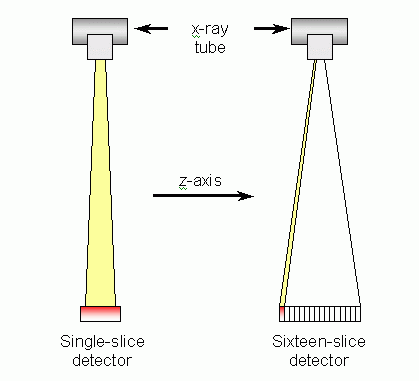
Figure 2: Single- and sixteen-slice detector scanners
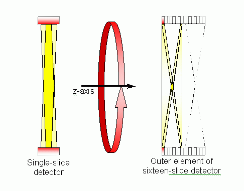
Figure 3: Cross sections through the volumes covered by rotation of single- and sixteen-slice scanners
The impact of cone beam artefacts can be reduced by using true three-dimensional reconstruction techniques, such as the Feldkamp algorithm, rather than the usual two-dimensional filtered back projection algorithm. The main drawback to the Feldkamp and modified Feldkamp techniques is that they are very computationally expensive, and take a long time to reconstruct. Other techniques that can be used include the ASSR (Advanced Single Slice Re-binning) algorithm which uses helical scan data to reconstruct multiple slices that are angled to the conventional transaxial scan plane (Figure 4). The angle of the image nutates around the transaxial plane, depending on the tube angle. These images can be filtered in the z-axis to produce conventional transaxial images.
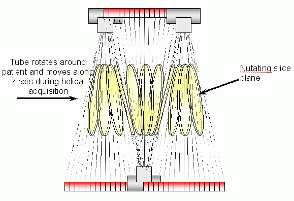
Figure 4: Slices produced using ASSR reconstructions
GE uses ASSR techniques for the LightSpeed Ultra. Siemens uses an algorithm known as AMPR (Adaptive Multi-Planar Reconstruction), an adaptation of ASSR that allows more flexible pitch options by using many partial- or multiple-rotation reconstructions at each scan position. Toshiba and Philips use adaptations of Feldkamp algorithms called ConeView and COBRA respectively.
Applications
The clinical applications featured at RSNA 2001 were built on areas that featured strongly in the scientific sessions. The issue of CT screening for both colon and lung cancer was discussed at length during the meeting in a number of sessions.CT imaging of the colon and lung is capable of producing high-quality images, which can help to identify early stage cancers. In order to demonstrate improved clinical outcomes for the patients involved, there is still a need for randomised clinical trials of screening programmes. Unfortunately, these sorts of exercises are both lengthy and complicated, and involve very large numbers of people. This has not stopped clinics in the US offering lung and colon screening services. Away from the controversies, one issue in colon screening that attracted considerable attention was data display techniques. Reporting times of 20 to 30 minutes per colonography exam are typical, so any method of reducing this time would be very valuable. The use of endolumenal views of the colon, known as virtual colonscopy, where the viewer examines the colon from inside and is able to 'fly' through, is an established viewing method (Figure 5). Modern workstations make this a more useful, interactive technique. Virtual colonscopy does have drawbacks: it cannot always show the entire colon wall, due to the bends and folds in the colon. A number of variations on this technique were demonstrated, including using simultaneous multiple views in different directions, and displaying a straightened, unfolded image of the colon.
Computer-aided detection is another area of interest for both colon and lung screening, drawing the attention to regions of the bowel or lung that the software assesses as being similar to a polyp or nodule.
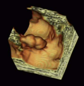
Figure 5: Endolumenal view of colon, showing large polyp (courtesy Voxar Ltd)
All the manufacturers of CT scanners demonstrated their CT perfusion packages. Perfusion imaging is mainly used to assess blood flow to the head, and can assess absolute and relative blood flow to different sections of the brain. Regions of interest are placed over the artery and vein.The same region of the brain is repeatedly scanned for a period of a ninety seconds to two minutes after the injection of contrast agent. As the contrast appears in the brain, the CT number changes over a period of time. From this data, regional blood flow can be measured. The results are generally displayed in images similar to Figure 6. Available parameters included are cerebral blood flow rates, mean transit times, blood volume and time to peak perfusion.
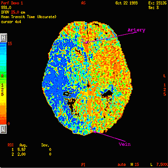
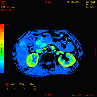
Figure 6: Typical CT perfusion images of the brain and kidneys (courtesy GE Medical Systems)
Potentially, perfusion imaging can also be used for other organs such as the liver, pancreas, kidneys or lungs. Most clinical packages do not include these yet. This is part of an interesting trend for CT scanners towards functional imaging.
Another organ where CT functional imaging has taken off is the heart. CT cardiac imaging was improved greatly by the introduction of multi-slice scanners, where scan data can be synchronised with the patient's ECG signal to provide effective scan times as short as 0.1 s. This enables the heart's motion to be frozen, and high-quality images of the myocardium and coronary arteries to be produced.All the scanner manufacturers have packages that use this technique. New this year at RSNA was the use of this data to calculate functional parameters related to the heart. The assessment of cardiac volume at different cardiac phases, ventricular ejection fractions, wall thickness and movement are all possible. Another area that shows promise is perfusion imaging of the coronary arteries. Although not yet clinically established, it can offer very high spatial resolution compared to nuclear medicine SPECT studies.
CT doses, particularly in children, were under focus at RSNA. GE and Siemens demonstrated their work on paediatric protocols. These provide methods for adjusting scan parameters correctly for the appropriate patient size. GE bases its system on a wider initiative, known as the Rainbow System, where children are divided up into nine different age and weight bands, and the bands colour coded accordingly. GE has protocols on its scanners optimised for each colour code. Siemens has a system that modifies the tube current based upon the patient weight for head exams and patient age for bodies.
With dosimetric issues in the spotlight, manufacturers had the opportunity to show their dose modulation systems. Last year, GE and Siemens had software that varied the current to the X-ray tube as it moved around and along the patient. The aim was to optimise the dose to the patient, increasing it for thicker cross sections and reducing it where it is not necessary. This has a number of benefits including potentially reducing patient dose, keeping image quality constant along the patient length, reducing tube loading and minimising streak artefacts caused by photon starvation. Toshiba announced a similar system for inclusion on its next multi-slice scanner software update, Real EC (Exposure Control). GE has now extended the system on its single- and dual-slice scanners to the LightSpeed series. This is known as OptiDose. Philips intends to introduce a system, DoseRight, to the Mx8000 in the near future. Siemens' CARE dose system is already available for its multi-slice scanners.
Future and summary
The logical endpoint of the technical advances discussed in this article lies in large volume scanners, such as Toshiba's 256 x 0.5 mm detector, or devices based upon flat panel detectors. These will be able to image entire organs in a single rotation. Multiple rotation imaging will improve functional studies, allowing observation of the behaviour of organs over time. We may start to see these research systems becoming a commercial reality in three to five years' time.
The move from single- to four-slice CT scanning that occurred three years ago opened up new clinical possibilities, such as cardiac imaging and improved angiographic and volume based studies. Eight- and sixteen-slice scanners offer potentially improved image quality but it remains to be seen how much additional diagnostic information they will provide. Whether the increased costs of eight- and sixteen- slice scanners over existing four-slice models can currently be justified in terms of benefit to patients is a debatable point.Their advantages lie mainly in better z-axis spatial resolution over four-slice scanners for protocols that cover extended scan lengths, providing image volumes with near-isotropic spatial resolution. They also further the paradigm shift that is occurring in CT scanning: from a two-dimensional imaging medium to one that isnatively three-dimensional.