:: rsna 2002: what's new in ct
This article was produced for the quarterly publication 'Diagnostic Imaging Review', printed and published by the Medical Devices Agency (now absorbed into the Medicines and Healthcare Products Regulatory Agency).
CT systems and hardware
Compared to its predecessors, 2002 was a year of relatively few new product announcements for CT at RSNA. The pace of change of the CT market since 1998, when modern multi-slice scanning was introduced, has been staggering, with current state of the art systems being 20 to 30 times more powerful than their single-slice counterparts. Within that four year period, GE for example, has introduced a new scanner at the top of its product range each year. It is therefore not so surprising that no major changes in the top of the range systems were announced this year.
The focus this year has been more orientated towards delivering upon the promises made last year, when the first details of the four main manufacturers' 16-slice scanners emerged. In addition, a number of gaps in the manufacturers' product line-ups have been filled. There is a suggestion that as the current systems are capable of performing nearly all the tasks required of them, the next step forward in design does not represent a huge improvement in clinical benefit over existing 16-slice systems. We may well see 32 or even 64-slice systems in the near future, but it might not be as simple to justify the benefits of these scanners over 16-slice systems as it was when comparing 16-slice to 4 or 8. One manufacturer said that they didn't necessarily believe that more slices brought huge benefits when compared to the extra cost, but as one of their competitors will bring a higher specification system out eventually, they must be ready to compete!
Towards cone beam scanning
The next major step forward in CT will come with volume CT scanners. These systems will be able to cover entire organs with a single rotation. All manufacturers are working on this type of system, although practical products are still a few years away. Toshiba showed their 256 x 0.5 mm row detector matrix, which covers a total length of 128 mm per rotation. GE talked in the scientific sessions about their system, which uses silicon flat panel imagers, similar to those used in planar imaging. There are many practical difficulties to be overcome with volume CT scanners, such as the data output (up to 1.5 Gigabytes per rotation in the GE system), handling and reconstruction. The main applications of these systems lie in areas such as perfusion imaging, where the uptake of contrast media can be tracked over a period of time. This is part of a general expansion of the capabilities of CT towards functional as well as anatomical imaging, for example, allowing assessment of cardiac function in terms of ventricular volumes and cardiac ejection fractions.
Benefits of 16-slice scanning
What benefits do the current state-of-the-art, 16-slice scanners provide for users? Table 1 shows the main scanner specifications
| Scanner | Main detector collimations (mm) | Tube heat capacity (MHU) | Max generator output (kW) | Fastest scan time (s) | Helical pitches |
|---|---|---|---|---|---|
| GE LightSpeed16 | 16 x 0.63, 16 x 1.25 | 6.3 | 54 | 0.5 | 0.562, 0.938, 1.375, 1.75 |
| Philips Mx8000IDT | 16 x 0.75, 16 x 1.5 | 6.5 | 60 | 0.42 | 0.5 - 1.5 |
| Siemens Sensation 16 | 16 x 0.75, 16 x 1.5 | 5.3 | 60 | 0.42 | 0.5 - 1.5 |
| Toshiba Aquilion 16 | 16 x 0.5, 16 x 1.0, 16 x 2.0 | 7 | 60 | 0.4 | 0.625 - 1.5 |
Table 1: 16-slice scanner specifications
Sixteen slice scanners are used in two main acquisition modes, providing either 16 sub-millimetre (0.5 - 0.75 mm) slices or 16 slices of double that width (1.0 - 1.5 mm) per rotation. In addition, the Toshiba scanner can also image sixteen 2 mm slices per rotation. These slice width values compare well with the pixel sizes of 0.5 and 0.8 mm at typical fields of view of around 250 and 400 mm for head and body imaging respectively. This means that they are capable of providing images with resolution that is identical or nearly identical in all three dimensions, which is also known as isotropic imaging. This is the ideal situation for any cross-sectional imaging modality and it is now possible to think of CT scanners acquiring volumes of data, which can be viewed in any plane, rather than a series of transaxial images.
The multi-planar nature of CT imaging was exemplified by Siemens' announcement of their asynchronic reconstruction algorithm, which will be available towards the end of 2003. This allows direct reconstruction of oblique images without the current intermediate step of reconstructing standard axial images.
Scan speeds for the 16-slice scanners are getting faster too. Rotation times of 0.4 seconds are available on the Philips, Siemens and Toshiba scanners. The main advantage of this is in cardiac imaging, where faster scan speeds allow patients with a wider range of heart rates to be imaged. The faster scan times are not generally used for conventional imaging modes, as they may involve compromises in spatial resolution due to a smaller number of views being acquired in the shorter rotation time. Routine scan times of 0.5 to 0.8 seconds tend to be used for body imaging in order to allow fast coverage of large scan volumes, and keep to a minimum the problems associated with patient breath-hold and contrast media equilibrium.
New scanners
In addition to the interest in the high-end systems, there were a number of multi-slice systems announced at intermediate points in the market. Siemens launched two scanners, the Somatom Emotion 6, and the Somatom Sensation 10. Marconi introduced the Mx8000IDT 10. Summaries of these systems are shown in Table 2.
| Scanner | Main detector collimations (mm) | Tube heat capacity (MHU) | Max generator output (kW) | Fastest scan time (s) | Helical pitches |
|---|---|---|---|---|---|
| Philips Mx8000IDT 10 | 10 x 0.75, 10 x 1.5 | 6.5 | 60 | 0.42 | 0.5 - 1.5 |
| Siemens Somatom Emotion 6 | 6 x 0.5, 6 x 1, 6 x 2, 6 x 3 | 4.2 | 40 | 0.8 | 0.5 - 1.5 |
| Siemens Somatom Emotion 6 Power | 6 x 0.5, 6 x 1, 6 x 2, 6 x 3 | 4.2 | 48 | 0.6 | 0.5 - 1.5 |
| Siemens Somatom Sensation 10 | 10 x 0.75, 10 x 1.5 | 5.3 | 60 | 0.5 | 0.5 - 1.5 |
Table 2: 6 and 10-slice scanner specifications
The Philips Mx8000IDT 10 and Siemens Sensation 10 use the same gantry and detectors as their 16-slice counterparts. The Siemens Emotion 6 is based upon a new detector design, shown in Figure 1, within the existing Emotion gantry.

Figure 1: Detector design for the Siemens Somatom Emotion 6
Another new scanner shown at RSNA 2002 was the GE e-Speed. This is a development of the electron beam tomography (EBT) technology, purchased when GE acquired Imatron in 2001. The e-Speed features a 1.5 mm slice thickness setting and a new, faster scan time of 33 milliseconds (0.033 s), compared with 3 mm slices and 50 milliseconds scan time on the previous EBT C300 scanner. In order to achieve this scan time, the maximum electron-beam current has been increased from 600 to 1000 mA.
PET-CT developments
New PET-CT systems were also on display, with scanners from GE, Philips, Siemens, and CTI Molecular Imaging. The Gemini system from Philips incorporates the dual-slice Mx8000D, whilst the GE Discover LS16, the Siemens Biograph Sensation 16 and the CTI Reveal XVI all feature 16-slice CT scanner elements. PET-CT is a clinical area that has taken off in recent years; the functional imaging provided by the PET images are complemented by the high spatial and contrast resolution of CT, allowing tumours to be localised accurately. In addition, the CT information can be used to perform attenuation correction of the PET scans. One of the main reasons that its use has expanded in the US is the availability of reimbursement from health insurers for a range of cancers. In the UK, the cost of the scanners and high running costs means PET, and PET-CT has a limited, but growing, market.
Applications
The hot issue in CT applications this year was CT screening examinations. The opening session of the scientific meeting was devoted to the topic, and it was referred to in a number of other talks.
CT Screening
There are a number of screening examinations that can be used on asymptomatic patients, although some of these patients may be within targeted high-risk groups. The main exams are colonoscopy, cardiac calcification scoring and lung cancer screening, as well as whole body protocols that are used as a general CT 'health check'. There are an increasing number of centres that are offering these services in the US. Privately run centres offering some of these examinations have also opened in the UK. NHS institutions do not generally provide CT screening examinations currently, but the issue is sure to become more important in the near future.
This issue is also important for manufacturers of CT scanners, as large scale screening programmes that could come out this work will require huge investment in equipment. There were a lot of new software tools for dealing with screening data, particularly for lung nodule identification and analysis (Figure 2), virtual colonoscopy and cardiac assessment. Particular emphasis was placed upon automation, so that organs or regions of interest within the scan volume could be isolated with the minimum amount of user intervention.

Figure 2: Volume rendered lung image showing nodule near chest wall. Image courtesy of TeraRecon, Inc
The opening scientific session was presented as a debate between two clinicians who were pro and against, CT screening.
The pro case is fairly simple - that by testing for diseases that kill many millions of people worldwide each year, screening examinations can catch the diseases in the early, pre-clinical forms. This should allow more effective treatment if the cancers are in stages that are less lethal. A prime example is lung cancer - when caught at stage I, when it can be resected, five-year survival rates are in the region of 60 to 70%. Overall lung cancer five-year survival rates for all stages of the disease are between 11 and 14%. Lung cancer screening trials among targeted groups, above the age of 65 and who have smoked the equivalent of one pack a day for 20 years, show lung cancer detection rates of about 0.5%. Similarly, the first sign of cardiovascular disease for more than 150,000 people per year in the US is sudden death. Calcium scoring of the coronary arteries has a good negative predictive probability - without calcified coronary arteries, the patient is unlikely to have coronary heart disease.
The case against CT screening is less straightforward. Although it is acknowledged that early detection of tumours will save lives, there are other effects to bear in mind. For any diagnostic case, there are four possible outcomes; true negative, true positive, false negative and false positive. True negative outcomes reassure the patient of their health. True positive results mean that disease may be found early, although not always early enough to affect clinical outcome. The problems come with the false results. False negatives can lead to a patient ignoring later signs and symptoms of disease, reassured by an incorrect diagnosis. False positives identify a disease that does not exist leading to follow-up diagnosis and treatment, each of which has an associated mortality, morbidity and cost. Another problem associated with screening programmes is 'overdiagnosis' when benign abnormalities that would not present themselves clinically are followed up. Again these follow-up procedures are not without risk or cost. In addition, there are radiation dose issues to consider.
The debate seems likely to continue in years to come. Action is being taken on the question of efficacy of lung cancer screening with the instigation of a multi-centre trial, sponsored by the US National Cancer Institute. This trial will look at 50,000 smokers, dividing them randomly into groups who will have CT screening or standard chest X-rays. The effect upon survival of the patients will be studied, in order to judge the usefulness of these techniques. The study is expected to last five to nine years, costing approximately $200 million. See http://cancer.gov/nlst/ for more details.
Patient dose and automatic exposure controls
Another issue that is assuming increasing importance in the US market is patient dose, particularly dose to paediatric patients. Although this topic has received closer attention in the UK and Europe in the past, the focus of the world's largest market for CT scanners tends to bring about action from the manufacturers.
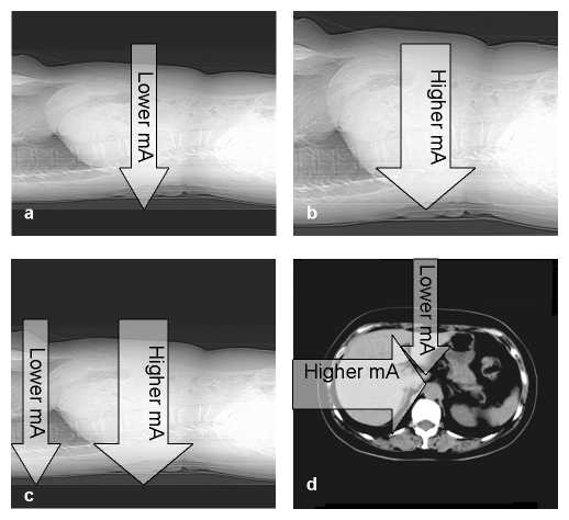
One of the best ways to optimise patient doses from CT is to have a system that varies the tube current (mA) according to patient attenuation. If the mA is adjusted relative to the patient thickness and density, the signal to the detectors remains more constant resulting in better image noise and patient dose characteristics. There are a number of ways to achieve this (see Figure 3):
- from patient to patient, so that smaller patients receive lower mA exposures than larger patients - patient size adapted mA
- along the patient's length (the scanner's z-axis), when areas of lower attenuation receive lower mA exposures than areas of higher attenuation. For example in an exam covering the chest and abdomen, the scanner uses a lower mA for the lung region than the abdomen - z-axis mA modulation
- during the course of a single rotation, so that lateral projections receive higher mA than the anterior-posterior projections, which are generally lower in attenuation - angular mA modulation
Overall, these techniques provide an equivalent role to an automatic exposure control (AEC) in general X-ray, but achieve it by varying the tube current rather than the exposure time. The systems may be controlled by reference to the patient attenuation from a localiser image (Scout View, Scanogram or Topogram), or by use of feedback from the previous rotation during a spiral scan run.

Figure 3: Varying patient attenuation and tube current for a) and b) patient size adapted mA, c) z-axis mA modulation and d) angular mA modulation
All manufacturers now have systems on their multi-slice scanners that use one or more of these techniques, aiming to ensure that the dose delivered to the patient is optimised. This has the additional advantage of providing a more consistent image quality from one patient to the next and/or along each patient's length. Streaking artefacts caused by photon starvation are less likely to occur when these systems are used, as the number of photons reaching the detectors is less variable. The systems (as of December 2002) are summarised in Table 3
| Manufacturer | Patient size adapted mA | z-axis mA modulation | Angular mA modulation |
|---|---|---|---|
| GE | AutomA | AutomA | N/A |
| Philips | DoseRight ACS | N/A | N/A |
| Siemens | N/A | N/A | CAREDose |
| Toshiba | N/A | RealEC | N/A |
Table 3. Summary of AEC systems for multi-slice CT scanners
GE's AutomA system provides patient size adapted mA, where the exposure is controlled by the prescription of a 'noise index' prior to scanning. AutomA also features z-axis dose modulation. This should be upgraded in 2003 to SmartmA, which also includes angular mA modulation.
Philips' AEC system is generally known as DoseRight, with DoseRight ACS (Automatic Current Selector) available currently. They are working on implementations of angular and z-axis mA modulation.
Siemens' CARE Dose is an angular mA modulation system, based upon feedback during helical scan runs. CARE Dose has the ability to adaptively reduce mA from a prescribed maximum value. A new version, available in 2003, should allow the increase in tube current where necessary. Siemens is working upon a patient size adapted mA system, based on a Topogram view.
Toshiba's package is known as RealEC (Real Exposure Control). This z-axis mA modulation system uses attenuation from a Scanogram image to define the tube current along the patient's length.
Conclusions
The 16-slice systems announced at RSNA 2001 are now a clinical reality, with hundreds of scanners installed worldwide by the end of 2002. They represent a potential for real improvements in a range of applications over the 4-slice models that have rapidly become established over the past four years. As the 16-slice detector banks are nearly the same total lengths, at between 20 and 32 mm, the maximum scan speed is not improved. The advantage lies in the ability to cover large volumes quickly, with excellent z-sensitivity. Isotropic imaging, where the resolution is approximately equal in all three dimensions, can routinely be achieved in CT now. The data that are gathered can be thought of as a volume, rather than a series of slices.
The screening of asymptomatic patients for a range of diseases has increased considerably in recent years. Real gains for the patient population have yet to be proven. The commencement of the US National Lung Screening Trial is particularly welcome to attempt to answer at least some of the many questions surrounding CT screening.
Patient dose is another controversial issue in radiology, and CT certainly contributes a very large proportion of the collective dose due to medical exposures. Automatic exposure control systems have existed on general X-ray equipment for many years, but were not implemented on CT scanners until recently. The currently available AEC systems for CT show promise. Future versions should help ensure that, wherever possible, dose from CT scanning is at its optimum level in order to produce acceptable diagnostic images.