:: rsna 2003
ImPACT was involved with a number of presentations and educational exhibition posters at the RSNA 2003 meeting in Chicago, which runs from the 30th November to the 5th December.
See this page for more details and pdf versions of the talks and posters, or read on for a report on the meeting.
CT Scanning at RSNA 2003
Introduction
RSNA 2003 saw the announcement of new scanners from all four main manufacturers. The systems include models that are capable of acquiring more than the current limit of 16 slices per rotation, and also units with extra-wide gantry bores for radiotherapy planning and scanning large patients. The new systems incorporate some new hardware components, including x-ray tubes and detectors.
Scanner enhancements
The headline announcements of the show included systems that acquired 32, 40 or 64 slices per rotation, but these works in progress are products that will not be available for purchase until around the time of RSNA 2004. There were, however, a number of new systems on show at RSNA 2003 that will be available in the shorter term.

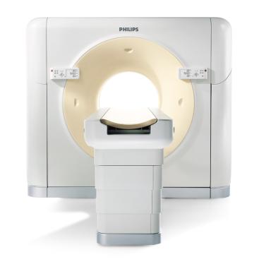
Figure 1: One of the models in Philips' new Brilliance range of CT scanners
Philips unveiled their new Brilliance range of scanners, which replace the Mx8000 range (Figure 1). The range includes 6, 10 and 16-slice scanners that directly replace the old Mx8000 equivalents. The scanners have a different look and now feature Philips' SureView reconstruction hardware. As standard, these systems can reconstruct 6.5 images per second, but there is the option to upgrade to 20 images per second. The 16-slice system also comes in a 'Power' configuration, which is capable of reconstructing 40 images per second, and uses the new MRC x-ray tube. Incorporating liquid metal spiral groove bearings, this tube has an 8 MHU heat capacity and a high cooling rate, producing an effective heat capacity several times higher than the nominal 8 MHU value. The MRC is Philips' own tube, developed from their image intensifier range. It incorporates a smaller focal spot than the tubes currently used in Philips systems, which will enable spatial resolutions as high as 24 lp / cm (MTF2). Other changes to the Philips range include a re-designed user interface, and the ability to initiate image reconstruction from remote workstations without having to return to the scanner console.
The LightSpeed Pro 16 from GE is an upgraded version of the existing LightSpeed 16. It is available in 60, 80 and 100 kW generator versions, and together with a new x-ray tube the system will be capable of scanning using at up to 800 mA at 120 kV, with a fastest gantry rotation time improved from 0.5 to 0.4 seconds.
A new addition to the Toshiba family of scanners is the Aquilion 8, based on the same hardware as the 16-slice system, but restricted to acquire 8, rather than 16-slices per rotation. The Aquilion 16 CFX is an enhanced version of the Aquilion 16, with a faster gantry rotation time of 0.4 seconds for cardiac applications.

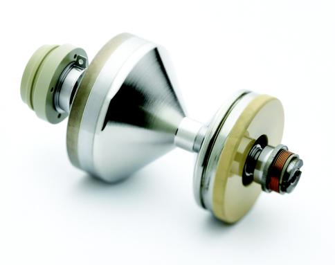
Figure 2: Siemens' Straton x-ray tube
One of the more interesting developments at RSNA was the Siemens Straton x-ray tube, which is currently available as an option on Sensation 16 scanners (Figure 2). The tube itself is a radical new design, where the entire tube body rotates, rather than just the anode, as is the case with conventional designs. This change allows all the bearings to be located outside the evacuated tube, and enables the anode to be cooled more efficiently. The Straton has a low inherent heat capacity of 0.8 MHU, but an extremely fast cooling rate of 5 MHU / min. This compares with typical figures of 7-8 MHU and up to 1.4 MHU / min for existing tubes. The heat capacity and cooling rate combine to produce a tube which Siemens claim is '0 MHU', implying that tube cooling considerations are a thing of the past. Sensation 16 scanners fitted with the Straton tube now have a fastest scan time of 0.37 seconds.
Wide bore scanners
When Philips introduced their ACQSIM CT scanner in 2000, they had the market for wide-bore systems to themselves. The wider scanner gantry opening and extended field of view of this system is beneficial in radiotherapy planning applications. Patients undergoing breast treatment in particular can be scanned in the standard treatment position, and the large field of view allows the whole patient contour to be incorporated in the treatment plan. Other applications include obese patients who may not fit within existing gantries, and trauma, where there may be other medical equipment around the patient that cannot easily be removed.
GE has released their LightSpeed RT scanner, which is the first four-slice wide bore system on the market. The RT is similar to the existing LightSpeed scanner range, but with a gantry bore that has been expanded from 70 to 80 cm, and a maximum reconstructed field of view that has been increased from 48 to 65 cm by the use of special reconstruction techniques. The other difference is that it has a fastest scan time of 1 second, compared with 0.5 for existing LightSpeed systems.
Siemens announced their SOMATOM Sensation Open, a works in progress that is based on their 16-slice Sensation 16 system, but comes with an 82 cm gantry bore. It is possible to reconstruct images with an 82 cm field of view on this system, giving excellent coverage of the patient periphery. Data outside the 50 cm central field of view is not reconstructed using conventional techniques, as the data in this region is incomplete. The resulting images are suitable for radiotherapy planning, or generating PET attenuation maps. Siemens are also marketing the scanner as being suitable for patients that are too large to fit through a conventional scanner gantry. The Sensation Open will use the Straton tube.
Although Toshiba don't have a specific wide-bore system on the market, their conventional scanners have the widest bore in their class, at 72 cm.
Beyond 16 slices
All of the manufacturers provided some details of their future systems with the capability of acquiring more than 16 slices per rotation. As with the step from four to sixteen slice systems, the goal is to improve z-axis resolution, give greater patient coverage per rotation and result in shorter total scan times.
Toshiba announced a 32-slice version of their Aquilion range of scanners. The detector in the Aquilion 32 is based on the unit found in the 16-slice model, which gives up to 32 mm z-axis coverage per rotation, but consists entirely of 0.5 mm rows rather than a combination of 0.5 and 1.0 mm rows. This can give 32 x 0.5 mm, or 32 x 1mm slices per rotation.
The 40-slice Philips Brilliance 40 is based around a new diode detector and MRC x-ray tube. The 40 mm detector can acquire 40 x 0.625 mm slices or 32 x 1.25 mm slices per rotation. The version of the RapidView reconstruction engine used in this system will be able to produce 40 images per second, which will keep reconstruction times for all scan runs to an acceptable level.
Siemens' SOMATOM Sensation 64 scanner is capable of acquiring 64 rows of scan data in a 0.37 second full gantry rotation. This is achieved using a 32-row detector, in conjunction with their Straton tube. The design of this tube enables z-axis oversampling to be carried out - effectively flying focal spot in the z-direction. Flying focal spot is a well-established technique that is currently used only in the x-y plane to double the amount of projection data that is gathered, increasing the spatial resolution of the system. Z-axis oversampling extends this to the third dimension, and significantly suppresses spiral artefacts as well as improving z axis resolution.
GE was less forthcoming than the other manufacturers about their future systems, preferring to focus upon their newer scanners that were available now. They did indicate that they are also working on a 32 slice system, which is understood to divide their current 16 slice detector into 32 x 0.625 mm sections.
Clinical need?
It is important to remember, in the midst of the numbers game that could be seen to have driven CT development over the last few years, that scanners with a greater number of slices need to offer clinical benefits in order for hospitals to justify their extra cost. As with going from four to sixteen slices, the changes that occur when going to 32 slices and beyond could be seen as incremental, rather than pushing back any great boundaries. The increased scanning speed and z-axis resolution of the new systems will probably be of most use in applications such as cardiology and angiography. In addition, perfusion imaging, which has yet to be established on a wide scale, will also benefit.
In cardiology, the entire heart can be imaged in a shorter time, which will lead to improved multi-sector reconstructions, as there is less time over which the patient's heart rate can vary. Alternatively, more phases could be used, giving better temporal resolution. Angiography will benefit from more coverage per rotation so the enhancement of vessels by contrast media will be more uniform. Toshiba showed some early clinical results from their 32 slice system, with a neurological examination that showed enhancement entirely within the arterial phase.
Future developments
During the scientific sessions Siemens presented data on the performance of a prototype system based on a flat-panel detector. With coverage in the z-axis of about 15 cm the system would lend itself to whole-organ imaging. The properties of the amorphous silicon detector produce images with excellent high contrast spatial resolution. Rotation times of the order of 20 seconds, and relatively poor low-contrast resolution due to the low signal to noise ratio mean that the applications of such a system in its current form will, however, be limited. Procedures that may benefit from this type of scanner are those such as surgical planning, where large coverage per rotation with high spatial resolution is useful. High radiation doses are required to improve on the low contrast resolution of the system.
On a similar theme, Toshiba showed work from a scanner based upon their 256 x 0.5 mm row detector, looking at issues relating to scattered x-rays along the z-axis, which will become more of an issue as the coverage per rotation increases.
Dose issues
GE, Siemens and Toshiba have expanded the capabilities of their automatic exposure control (AEC) systems, which work by varying the x-ray tube current in response to changes in the patient's attenuation.
The Toshiba system, now known as SUREExposure, has been expanded, and the operator can request a desired image quality by selecting an image noise level. The system then sets the appropriate mA for each rotation in order to achieve this image quality.
The Siemens CAREdose system is now capable of modulating the tube current along the length of the patient, and also accounts for overall patient size. CAREdose 4D takes a typical mAs value used for a standard sized patient as the starting point for modulation, and adjusts this based upon the patient size This is in addition to the rotational modulation of the mA that was available in existing versions of CAREdose.
GE's SmartScan system has been expanded on the LightSpeed Pro 16 to allow rotational mA modulation, as well as the existing z-axis and overall patient size adjustment that is controlled by setting a desired image noise level.
PET-CT
PET-CT is now established well clinically, with hundreds of systems installed worldwide. The relative costs of a stand-alone PET system compared to a combined PET / CT scanner mean that buying a stand-alone system is no longer a sensible option when the benefits that CT brings are considered.
New systems announced at RSNA include a new 6-slice version of Siemens' Biograph PET / CT range to accompany the existing 2 and 16-slice versions. The PET component of all the Biograph systems is based on high-performance LSO detectors.
Philips' new PET / CT system is based around their 16-slice Brilliance CT scanner and an Allegro PET system with GSO detectors. The open design of the Philips system allows clinicians access to the patient throughout the imaging procedure, and reduces claustrophobia.
The second-generation design of the GE Discovery ST PET / CT system is capable of acquiring in 2D and 3D modes, and has a patient-friendly 70 cm bore.
ImPACT posters and presentations

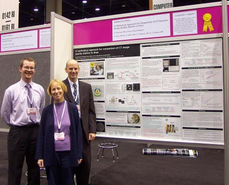
Figure 3: ImPACT members with one of their posters at RSNA 2003
ImPACT had two posters displayed at RSNA. The first, "Methodical approach for comparison of CT scanner image quality relative to dose", was awarded a certificate of merit (Figure 3), and the journal 'Radiographics' has asked that our second poster, "Artefacts in CT: recognition and avoidance", be submitted to them for publication. Work was also presented in two of the scientific sessions, "Low contrast detail detectability measurements on multi-slice CT scanners", and "A visual method for demonstrating the relative performance of cone beam reconstruction". Both talks and posters are available on-line.
Summary
2003 was an exciting year for developments in CT at RSNA. There were a number of changes to existing systems to expand their capabilities in areas such as faster scan speeds and higher tube output. The market for wide gantry bore systems which was started by Philips three years ago is now being competed for by other manufacturers, and is likely to expand with the availability of large field of view multi-slice systems.
The developments that really had people talking, however, were the previews of future systems capable of acquiring more than 16 slices per rotation. The initial clinical images looked very impressive, but some of the technical details were still a little unclear. 2004 should be another interesting RSNA from the perspective of CT, when these scanners become a commercial reality.