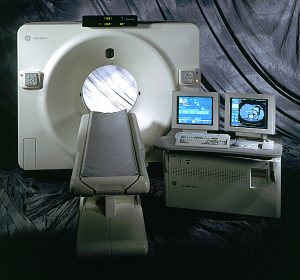
:: rsna 1999
Introduction
After a number of fairly quiet years in CT scanner technology, the last two years at RSNA have produced a lot of interesting developments. The announcement in 1998 of multi-slice detector systems, capable of simultaneously acquiring more than one CT image has been followed by further development of these units, most of which were software based. A large number of CT related presentations were also given in the RSNA scientific meeting, explaining some of the technology and applications that were originally announced last year, but which were not all fully implemented at the time. A number of single (and dual) slice scanners were introduced this year, and the possibilities of true volumetric CT imaging were starting to be explored, making this one of the busiest years for CT at RSNA ever!

View of Chicago from RSNA conference centre
Multi-Slice
Last year was the breakthrough for multi-slice CT imaging, with four manufacturers announcing multi-slice scanner systems (GE with the LightSpeed QX/i, Picker, who were re-launched this year as Marconi Medical Systems with the MX8000, Siemens with the Plus 4 Volume Zoom and Toshiba with the Aquilion). Although these scanners were introduced at the same time, they were at varying stages of development, and were not all ready for the wider market. This has changed in the past year, and all of these scanners are now in clinical use in European sites, and all but the Aquilion have been installed in the UK. Toshiba and Siemens also announced new multi-slice scanners.
Toshiba have added multi-slice capabilities to their existing Asteion VR scanner, known as Asteion Multi. This is offered either as a direct purchase, or an upgrade to an existing scanner. The upgrade involves the addition of the same multi-slice detector bank as is used in the Aquilion multi-slice scanner, which consists of 30x1 mm detector elements in the z-direction (the patient's long axis), with 4x0.5 mm elements at the centre, making a total z-thickness of 32 mm. These detectors allow combinations from 4x0.5 mm, through 4x1, 4x2, 4x3, 4x4, 4x5, 4x8, to 2x10 mm simultaneous slice acquisitions. A new computer system for the scanner is also included in the upgrade. This is the same unit used in the Aquilion, with 1 GB of memory and a 45 GB hard disk. The main differences, therefore between the Aquilion and Asteion multi-slice systems are the scan speed (0.5 vs. 0.75 seconds), generator power (60 vs. 48 kW) and tube heat capacity (7.5 vs. 4 MHU). Toshiba see the Asteion Multi as a general purpose multi-slice scanner, with the Aquilion as the premium model, capable of the speed necessary for cardiology examinations.
Siemens have announced their new Volume Access scanner which should be available for purchase soon, based upon their Volume Zoom architecture, but capable of acquiring only two, rather than four simultaneous slices. It will be upgradeable to four slice scanning. Their new Emotion scanner (see Single Slice section) can also be upgraded to dual slice scanning.

The GE LightSpeed 4 slice scanner
GE have made some improvements to their LightSpeed QX/i scanner with the introduction of a dynamic beam collimator that is capable of adjusting its position every 20 ms, thus reducing the irradiated slice width. Dose overlaps that were 8 mm are now approximately 2 mm. For example, the irradiated width of a 4x5 mm slice, which was previously 28 mm, is now 22 mm.
Another improvement, soon to be introduced on installed LightSpeed scanners, and standard on new models, is sub-millimetre (approximately 0.6 mm) slice width scanning. This will be achieved through deconvolution of data acquired with a small pitch, rather than using a narrower detector width. GE also announced their 'Direct 3D' system which builds 3D images of the scan volume on screen as the scan takes place (within about 0.1 second of each slice being reconstructed). This can be advantageous in multi-slice scanning, as the large number of images produced by the scanners can make reviewing them a time consuming business, and viewing them directly in 3D can potentially increase viewing efficiency.
Marconi have integrated their Mx8000 scanner into their Venue interventional system, including their FACTS flat panel II, and PinPoint stereotactic system. This combined system is known as LifeFlight, and is being targetted for use in trauma suites, for diagnostic and interventional use.
Cardiac imaging dominated applications for multi-slice scanning, and is now a reality with fast, narrow slice acquisitions, combined with ECG gating. The speed of multi-slice scanners permits scans with a greater temporal resolution than was previously achievable, which reduces blurring due to cardiac motion. This speed is being realised by two different methods; prospective and retrospective gating.
- Prospective gating is where the ECG signal is used to trigger data acquisition in a user defined phase of the cardiac cycle, usually diastole. With a half scan reconstruction, (one that includes only 180° of data), a temporal resolution of approximately 0.3 seconds is achieved for a 0.5 second rotation scanner.
- Retrospective is gating, where scan and ECG data are acquired simultaneously such that for each slice, each detector will acquire data in the same phase of the cardiac cycle, but at a different angular position.

Volume rendered CT angiography of coronary arteries (Marconi Mx8000)
Combining data from subsequent heart beats into a single slice gives a potential temporal resolution of about 0.1 seconds for half scan reconstruction on a 0.5 second scanner. Acquiring data in this way results in a 4-D (time varying) image set.
Visualisation of the coronary arteries, and calcification scoring for assessment of stenoses are the main uses for these images. All of the manufacturers offer their versions of these applications. Marconi also offer their cardiac calcification package for their single and dual slice scanners.
Single Slice
With all the attention being focussed on multi-slice scanners, the manufacturers could be forgiven for standing still with their single slice models, but this was not the case, as a number of new scanners were announced this year at RSNA. It is interesting to note that it is ten years since the introduction of spiral CT scanning, and all scanners on the market that now offer this capability.
Philips have renewed their top of the range scanners, with the launch of the Secura and Aura systems. The Secura is the higher specification scanner with 0.7 second scanning (see table 1 for more details). Both scanners come with Philips' new scanner interface. This features 'WorkWise', a dual monitor and keyboard arrangement, with one of the monitors controlling the scanner and viewing images, and the second acting as a workstation style viewer.
Also new from Philips is their 'DoseRight' system that varies the x-ray tube current during acquisition to give a constant image quality. The current variation can be carried out over the course of a rotation, to account for different patient thickness for different projection angles, or within the course of a scan series to adjust for changes in patient anatomy e.g. when moving from the abdomen to the chest. A tube current adjustment for overall patient size can also be made. These tube current calculations are made from the scanogram images.
Siemens showed their new Emotion and Balance scanners, which are general-purpose 0.8 and 1 second scanners respectively. These are replacing the Plus 4 Expert systems in the Siemens product range. They will be upgradeable to dual slice scanning. Current models are operated using a similar Unix based system, as on the Plus 4, but a new interface based on Siemens' new 'Syngo' platform, as seen on the Volume Zoom multislice scanner, will be fitted to current models when it is available in the near future. Syngo is the Windows NT based interface that Siemens are now using on all their new digital systems. It aims to standardise the control of digital imaging devices, reducing the need for retraining and improve ease of use.

The Siemens Emotion scanner
GE also announced two new scanners, the CT/e and ZX/i. The CT/e is a compact, basic scanner offering helical scanning with solid state detectors. The interface is the same as on the /i range, but runs on a Silicon Graphics O2 computer, rather than the more powerful Octane. The ZX/i tops the FX/i and LX/i range, offering a marginally faster 0.7 second scan, using the Solarix 630 metal x-ray tube. Shimadzu demonstrated a works in progress version of their SCT-7800TX scanner, which should be available later this year, which features a 0.75 second scan time and a compact installation.
| Manufacturer | Model | Fastest Scan Time (s) | Tube Heat Capacity (MHU) | Generator Output (kW) | Gantry Aperture (cm) |
|---|---|---|---|---|---|
| GE | CT/e | 1.5 | 2 | 24 | 65 |
| GE | ZX/i | 0.7 | 6.3 | 53 | 70 |
| Philips | Aura | 1.0 | 3.5 | 30 | 67 |
| Philips | Secura | 0.7 | 7.7 | 60 | 72 |
| Shimadzu | SCT-7800TX | 0.75 | 3.5/4 | 48 | 70 |
| Siemens | Balance | 1.0 | 2 | 26 | 70 |
| Siemens | Emotion | 0.8 | 3 | 40 | 70 |
Table 1. Summary of new single slice scanners at RSNA 1999
Other CT scanner Issues
Hitachi introduced a new rotational angiography system, the SF-VA 100, which is capable of reconstructing 3-D images from its full rotation acquisitions. It consists of a 12 inch image intensifier system enclosed in a CT style gantry with a 70 cm aperture. The image intensifier acquires at 60 frames per second, over a 2.7 (180º) or 5.4 (360º) second acquisition. The entire volume of data acquired can then be reconstructed, and viewed as a volume rendered, surface rendered or MIP rendered image. They stress that this is not a CT scanner, but for truly isotropic volume imaging (i.e. resolution in z-axis is the same as x and y-axes), this looks to be an interesting development. GE also showed work in progress of 3-D reconstructions from a rotating flat panel detector, acquiring projections at 30 per second.
Aside from cardiac imaging, the other hot clinical topic is the use of CT in low dose screening for lung cancer. The Early Lung Cancer Action Project (ELCAP) developed at Cornell and New York University, and trialled also at a number of other centres, screened 1000 smokers over the age of 60 for lung nodules using a low dose procedure (140kV, 40mA, pitch 2 on a GE HiSpeed Advantage). Malignant nodules were found in 37 patients within this group, but the encouraging statistic is that 80% of these nodules were at stage 1A in their development, catching them much earlier than is currently possible. This is important as currently, lung cancer has a 10-15% 5 year survival rate, whereas for cancers caught early, survival rates have been estimated in the region of 80%.
There is still a lot more work to be done before lung screening becomes routine, including follow up studies to determine the true survival rates. More details can be found at http://icscreen.med.cornell.edu