:: rsna 2000 part 2: applications
<<< part 1: introduction and hardware
Cardiac CT
Of all the applications that have benefited from the recent advances in CT technology, it is cardiac imaging that is currently receiving the most attention. Judging from the papers in the scientific sessions at RSNA, it is also an area that the manufacturers are devoting a lot of their development efforts to.
Conventional single slice CT scanning has a number of drawbacks in relation to imaging the heart. Firstly, the scan times of approximately 1 second mean that cardiac motion blurs the image; secondly, in order to get reasonable coverage in the z-axis during one breath hold, the imaged slice thickness has to be compromised.
Electron beam CT (EBCT) scanners, produced by Imatron, do not suffer from these problems, as they have a scan time of 0.1 seconds and multiple target and detector rings for improved z-axis coverage. They have therefore been the cardiac CT imaging device of choice until recently, but are not as widespread as conventional CT scanners, as they are not as versatile and are more expensive to purchase and operate.
Modern multi-slice CT scanners have made considerable advances in improving on the performance of single slice scanners by using the multiple detectors to produce a composite image that improves the temporal resolution, and have fast rotation speeds that increase the coverage during a single breath hold.
There are two general approaches to acquiring cardiac images on multi-slice scanners, prospective and retrospective gating. Both use an ECG wave to 'gate' or synchronise scan data with the cardiac cycle.
Prospective gating triggers the start of axial scanning to a chosen point of the R-R interval in the ECG cycle (see Figure 1). This will be chosen to correspond to the region of the cardiac cycle where cardiac motion is at a minimum. Half scan techniques (which need only 180° of data, gathered from approximately 240° of a gantry rotation), can give a temporal resolution of approximately 0.3 seconds for a scanner with a 0.5 second rotation time.

Figure 1 ECG signal, R-R interval and Prospective
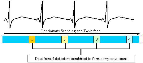
Retrospective gating (see Figure 2) uses continuous scanning, at an x-ray beam pitch of approximately 0.25 (image pitch of 1) at a fast rotation speed. The ECG data that has been simultaneously acquired can be used to reconstruct the correct scan data in a similar manner to the prospectively gated method. In addition, a so-called 'multi sector' or 'multi phase' reconstruction can be performed, where data from more than one detector, at different projection angles, but similar heart phase is combined to produce composite scan projection sets. For four slice scanning, this can improve the temporal resolution by up to a factor of four relative to prospective gating, and a temporal resolution of approximately 0.1 seconds can be achieved, similar to that of EBCT.

Figure 2. Retrospective gating cycle
There are technical difficulties associated with both methods, particularly in relation to fast and variable heart rates. In retrospective gating, a resonance situation can occur, where the gantry rotation speed can match the patient's heart rate, and each detector views each portion of the heart at a similar cardiac phase. This particular problem can be partially overcome by varying the gantry rotation time, and this is the intention behind GE's 0.5, 0.6 ... 1.0 second scan times. Posters in the scientific exhibition at RSNA also discussed other manufacturers' scanners operating at different scan times.
Cardiac calcification scoring
One of the applications of cardiac CT scanning is the imaging of calcified vessels, which are a risk factor for coronary artery disease. The calculation of the volume of pixels above a certain threshold (usually 130 HU) enables the assessment of the degree of cardiac calcification. All the current multi-slice manufacturers except have calcification scoring packages. A number of centres presented work comparing the results of multi-slice cardiac scoring with EBCT scores. In general, although the multi-slice scanners produced different Agatson scores than EBCT, there was good correlation between the two, with high repeatability for multi-slice. Patient doses for EBCT were shown to be considerably lower than multi-slice, however, this seemed to be as much a result of the acquisition parameters as the inherent differences between the scanners.
Coronary angiography
Retrospectively gated cardiac imaging with contrast media makes the imaging of the coronary arteries practical, allowing non invasive detection of stenosis and visualisation and differentiation of soft plaque. A number of papers were presented on this subject in the scientific meeting, and although the results were generally favourably received, concerns remain about the visualisation of small vessels (less than 1 mm in diameter), and the ability to detect soft plaque in the presence of calcifications.
Dose Optimisation Systems
Three manufacturers presented their dose reduction and optimisation systems at RSNA, although only GE currently have a product in widespread clinical use, with Siemens and Philips expecting to introduce their system next year.
GE's SmartmA system is currently only available for their HiSpeed X/i series (not the LightSpeed), and adjusts the tube current relative to both the patient size, and along the z-axis. It uses AP and lateral SPR (scout) views to assess the attenuation of the patient at each z-axis position. Based on this information, relative tube current settings for each helical rotation are calculated, and the system can be operated in one of three ways. Firstly, the mA for each rotation can be scaled relative to a maximum value, secondly three totally automatic image quality levels (low dose, standard and image quality) requiring no manual mA setting can be selected, and finally, there is a clipping mode, where the automatic mA values are restricted to remain below a maximum value entered by the operator. SmartmA is claimed to ensure constant image quality in structures that vary in attenuation along their length (e.g. the lungs), and optimal use of patient dose. In addition, it potentially increases the coverage length for scans limited by tube heating considerations.
Siemens described their Care Dose optimisation system for their multi-slice scanners, which adjusts the tube current in the course of each rotation according to the attenuation at each scan angle. This information is gained from data acquired during the previous rotation, and therefore does not require extra SPRs. For areas such as the pelvis and shoulders, the attenuation in the lateral direction is much greater than the AP or PA, so this system ensures more constant noise levels in each scan projection, reducing streaking artefacts, and ensuring a more constant dose distribution throughout the patient's cross section. mAs reductions were reported as being typically 15-55%, depending on anatomical region.
Philips will soon be introducing their DoseRight system that is capable of adjusting to individual patient size, using a single SPR to suggest an mA for each study. Further developments to the system are planned to modulate the mA along the patient's length.
Other Developments
CT Perfusion
Another application that was being featured by all manufacturers was CT brain perfusion imaging. By continuously scanning during contrast injection, the change in CT number reflects the blood supply to different regions of the brain. Parameters such as cerebral blood flow and volume, mean transit time, time to start and time to peak can be calculated, and are particularly useful in the assessment of stroke patients.
Multi-modality imaging
The use of CT scanners to provide accurate anatomical data for PET and SPECT scanning was a feature of RSNA 2000. Work in progress from Siemens, GE and Philips/ADAC showed two-in-one systems that allow fusion of the high spatial resolution CT images with the functional information provided by nuclear medicine images.
A CT scanner tube and detector are also included in GE's Hawkeye gamma camera to provide attenuation correction data.
Lung Cancer Screening
CT lung cancer screening research is continuing, particularly in the US and Japan, following its first appearance at RSNA last year as a hot topic. Areas that were reported on included computer aided detection of nodules and optimisation of scan protocols, as well as cost effectiveness evaluations. The potential implications in terms of expense and workload of carrying out lung cancer screening on a wide basis means that this topic is sure to receive considerable attention in the future.
Conclusion
When modern multi-slice scanners were introduced at RSNA in 1998, very few systems were in clinical use. The intervening two years have seen many new installations, which has increased the experience of both manufacturers and users. In addition, the software applications promised at launch have begun to be employed in routine practice. Development is still continuing, and the goal that all companies are pursuing is isotropic imaging of larger volumes. This will change the emphasis of CT scanning from two dimensions to three and lead to a more accurate representation of human anatomy.
In the meantime, the continued development of applications such as cardiac, oncology and lung imaging, as well as functional imaging means that CT scanning is moving forward rapidly in a wide range of fields.